Key (online version)
|
|
1a |
Wings absent.
Included:
Ocydromiinae: Apterodromia Oldroyd, 1949 |
|
|
| b |
|
2 |
| |
|
2a |
| Wings modified in such a way that venation is incomplete or invisible. |
|
|
| b |
| Wings fully developed; if shortened then venation appears to be normal. |
|
3 |
| |
|
3a |
| Cell dm (discal cell) absent, vein M unforked AND stigma absent; R1 usually not reaching to the second half of the wing. Anal cell (cell Cu) usually absent; if present, then only very small and anal vein scarcely visible. Small predaceous species, 1-5 mm in length. |
| Tachydromiinae |
|
4 |
| b |
| Cell dm (discal cell) present; if absent, then EITHER vein M forked into M1 and M2 (Bicellaria [Fig. 282]) OR stigma visible, vein R1 reaching costa at about 1/3 from wing tip and vein CuA1 strongly recurrent (Leptopezella [Fig. 322]). Anal cell present. |
|
13 |
| |
|
4a |
| Postpronotum more or less differentiated, often sharply so. Eyes bare; head with distinct neck, not closely set upon fron of thorax. |
|
5 |
| b |
| Postpronotum not differentiated. Eyses microscopically pubescent; head without distinct neck, set closely upon thorax. |
|
9 |
| |
|
5a |
| Cell cup (anal cell) present, even though vein A1 (anal vein) may be very indistinct. Postpronotum rounded or not very long. Wings clear or indistinctly clouded. |
|
6 |
| b |
| Cell cup (anal cell) absent because vein A (anal vein) is missing (vein CuA2 may be present). Postpronotum conspicuously elongated. Wings distinctly clouded or banded. |
|
8 |
| |
|
6a |
| Eyes touching on frons. Wings large, narrow at base, broadened apically and blunt-tipped. Legs with all femora equally slender, without raptorial modification. |
| Symballopthalmus Becker, 1889 |
|
|
| b |
| Eyes at least very narrowly separated on frons. Wings with more or less developed axillary lobe, not conspicuously broader at tip. Legs usually more or less thickeend on anterior four femora and often with special raptorial modifications. |
|
7 |
| |
|
7a |
| Eyes at least very narrowly separated on facial part below antennae. Humeri not conspicuously large. Mid femora usually thickened; mid tibiae either with or without apical spur. |
| Platypalpus Macquart, 1827 |
|
|
| b |
| Eyes touching below antennae. Humeri very large. Fore femora the stoutest; mid tibiae always without apical spur. |
| Dysaletria Loew, 1864 |
|
|
| |
|
8a |
| Vein CuA2 (vein usually closing anal cell) present; wings usually uniformly dark clouded. Occiput below neck with conspicuous, whitish, bristle-like setae. |
| Tachypeza Meigen, 1830 |
|
|
| b |
| Vein CuA2 (vein usually closing anal cell) absent; wings usually banded. Occiput below neck with usual hairs only. |
| Tachydromia Meigen, 1803 |
|
|
| |
|
9a |
Cells br and bm (basal cells) equal in length. Face usually broad and often with deep jowls below eyes.
Many coastal species. |
| Chersodromia Walker, 1849 |
|
|
| b |
| Cell br (first basal cell) distinctly shorter than cell bm (second basal cell). Face linear or eyes practically touching below antennae. |
|
10 |
| |
|
10a |
| Veins R1 and R2+3 short, the latter ending in the costa at about middle of wing; wings short and often narrowed. Frons rather broad, parallel-sided. |
| Stilpon Loew, 1859 |
|
|
| b |
| Veins R1 and R2+3 longer, the latter ending in the costa beyond middle of wing; wings not conspicuously shortened or narrowed. Frons narrow in front, more or less broadened toward vertex. |
|
11 |
| |
|
11a |
| Jowls linear. Anterior pair of ocellar setae present. Base of Rs rather long (almost twice as long as length of R4+5 to junction with vein r-m). |
|
12 |
| b |
| Jowls deep below eyes. Anterior pair of ocellar setae absent. Base of Rs rather long (about as long as length of R4+5 to junction with vein r-m). |
| Crossopalpus Bigot, 1857 |
|
|
| |
|
12a |
| Hind tibiae with distinct long anterodorsal setae. Species yellowish in ground colour. |
| Elaphropeza Macquart, 1827 |
|
|
| b |
| Hind tibiae without distinct long setae. Species dar brown to blackish in ground colour. |
| Drapetis Meigen, 1822 |
|
|
| |
|
13a |
| Anal cell large, as long as or longer than second basal cell. Discal cell emitting two veins only, vein M2 absent. Hind femora more or less enlarged and/or with long ventral setae. |
| Hybotinae |
|
14 |
| b |
| Anal cell much shorter than second basal cell. Discal cell emitting three veins, though in Ocydromiinae M1 abbreviated or absent. Hind femora slender; if stout (Oedalea), then small ventral spines only near apex and discal cell emitting three veins. |
| Ocydromiinae s.l. |
|
18 |
| |
|
14a |
| Anal cell short, as long as basal cells; radial sector long, much longer than the distance from humeral crossvein to its origin. Wings maculated (in European species) [Fig. 278]. |
| Syneches muscarius (Fabricius, 1794) |
|
|
| b |
| Anal cell much longer than basal cells (in European species); radial sector rather short, at most as long as the distance from the humeral crossvein to its origin. Wings clear or uniformly clouded, not maculated [Fig. 255]. |
|
15 |
| |
|
15a |
| Basal sector of vein M (separating basal cells) scarcely visible, radial sector very short [Fig. 275]. Antennae inserted above middle of head in profile [Fig. 277]. Hind tibiae at tip and hind metatarsi thickened [Fig. 276]. |
| Syndyas nigripes (Zetterstedt, 1842) |
|
|
| b |
| Basal section of vein M (separating basal cells) distinct, radial sector rather longer [Fig. 255]. Antennae inserted at or below middle of head in profile [Fig. 252]. Hind tibiae and metatarsi simple. |
| Hybos |
|
16 |
| |
|
16a |
| Mesonotum polished black except for grey dusted narrow lateral and median strips. Fore and mid legs yellow. Male: fore tibiae and tarsi and hind legs with long pale hairs; genitalia rather small. |
| Hybos femoratus (Müller, 1776) |
|
|
| b |
| Whole surface of mesonotum thinly grey or brownish dusted, at most subshining. Fore and mid legs blackish. |
|
17 |
| |
|
17a |
| Large thoracic setae yellowish. Mid femur with a row of ad setae, mid tibiae with several strong av setae. Mesonotum finely brownish dusted. Hind metatarsus only with short ventral setae [Fig. 250], and at least anterior metatarsi translucent brownish. Male: fore tibiae and tarsi rather short pubescent; genitalia very large [Fig. 249]. |
| Hybos culiciformis (Fabricius, 1775) |
|
|
| b |
| Large thoracic setae black. No ad setae on mid femur and no strong av setae on mid tibia. Mesonotum very thinly grey dusted, rather subshining. Hind metatarsus with short but strong ventral spines [Fig. 256] and legs quite black. Male: for tibiae and tarsi with very long black hairs; genitalia scarcely broader than tip of abdomen. |
| Hybos grossipes (Linnaeus, 1767) |
|
|
| |
|
18a |
| Vein Rs short, arising near apex of cell bm. |
|
19 |
| b |
| Vein Rs long, arising before or in the middle of cell bm. |
|
21 |
| |
|
19a |
| Cell dm (discal cell) absent; vein M1+2 evanescent [Fig. 322]. |
| Leptopezella Sinclair & Cumming, 2007 |
|
|
| b |
| Cell dm (discal cell present). |
|
20 |
| |
|
20a |
Cell dm long; wing narrow and anal lobe undeveloped;
hind tibia bristled and not geniculate at base; hind femur
banded (Chile, Australasia). |
| Hoplopeza Bezzi |
|
|
| b |
Cell dm short; wing broad and anal lobe developed; hind
tibia lacking bristles and generally geniculate at base;
hind femur not banded (Chile, Australia). |
| Scelolabes Philippi |
|
|
| |
|
21a |
| Cell dm (disal cell) emitting three veins that reach the wing margin or cell dm absent but three medial veins reaching wing margin. |
|
22 |
| b |
| Cell dm (discal cell) emitting no more than two veins that reach the wing margin, the stump of a third vein may be present but that does not reach the wing margin. |
|
27 |
| |
|
22a |
| Cell dm (discal cell) absent [Fig. 282]. |
| Bicellaria Macquart, 1823 |
|
|
| b |
| Cell dm (discal cell) present. |
|
23 |
| |
|
23a |
| Fore femur greatly swollen, with 2 long basal spine-like setae; antenna inserted above middle of head. |
| Apteromyia Oldroyd, 1949 (winged species) |
|
|
| b |
| Fore femur slender; antenna inserted about middle of head. |
|
24 |
| |
|
24a |
| Species from South America [Male: Distiphallus long and flexible, nearly extending the length of the shaft.] |
| Neotrichina Sinclair & Cumming, 2000 |
|
|
| b |
| Not in South America. [Male: Distiphallus short, not nearly extending the length of the shaft.] |
|
25 |
| |
|
25a |
| Hind tibia slender. Wings stigma not extending to tip of vein R2+3 [Fig. 281]. At least three pairs (6-10) of scutellar setae. |
| Trichinomyia Tuomikoski, 1959 |
|
26 |
| b |
| Hind tibia dilated towards tip. Wings stigma extending to tip of vein R2+3 [Fig. 280]. Usually only two pairs of scutellar setae rarely 6-8 . |
| Trichina Meigen, 1830 |
|
|
| |
|
26a |
| Larger, body 2.2-3.1 mm, mesonotum subshining black due to a weak covering of browsnish dust, legs yellowish. Style as long as 3rd antennal segment. Female: frons very narrow, halteres light brown. |
| Trichinomyia flavipes (Meigen, 1830) |
|
|
| b |
| Smaller, body 1.6-2.2 mm, mesonotum polished black, legs blackish. Style much shorter than 3rd antennal segment. Female: frons polished black and verty broad, halteres blackish. |
| Trichinomyia fuscipes (Zetterstedt, 1838) |
|
|
| |
|
27a |
| Eyes with ommatrichia. |
| Abocciputa Plant, 1989 |
|
|
| b |
|
28 |
| |
|
28a |
| Abdominal sclerites heavily sclerotized, punctate; pulvilli lacking; male terminalia hidden inside segment 8; antennal style present (Australasian, Neotropical, Oriental and Palaearctic Regions). |
| Chvalaea Papp & Földvári, 2001 |
|
|
| b |
| Abdominal sclerites not heavily sclerotized; pulvilli present; male terminalia exposed; if the abdominal sclerites are heavily sclerotized then antennal style lacking (some Oropezella). |
|
29 |
| |
|
29a |
| Short M1 vein arising from cell dm. |
|
30 |
| b |
|
32 |
| |
|
30a |
Antennae inserted above middle of head. Anal lobe of wing weakly developed.
[According to Sinclair & Cumming (2000) this genus includes L. bimaculata (Bezzi, 1904) and several undescribed species, all from Australia.] |
| Leptodromia Sinclair, 2000 |
|
|
| b |
| Antennae inserted at about middle of head [Fig. 260]. Anal lobe of wing well developed [Fig. 259]. |
|
31 |
| |
|
31a |
| Larger, 4-5 mm in length. Palpi yellow, thorax black in male, yellow in female; acr 8- to 10-serial, short and pale. Male: halteres pale, abdominal pubescence whitish. |
| Leptopeza flavipes (Meigen, 1820) |
|
|
| b |
| Smaller, about 3 mm in length. Palpi blackish, thorax black in both sexes; acr 6-serial, black, very long in male, short in female. Male: halteres dark, abdominal pubescence blackish. |
| Leptopeza borealis Zetterstedt, 1842 |
|
|
| |
|
32a |
| Anal lobe not developed, wing narrow; in doubtful cases antennal inserted high on head. |
|
33 |
| b |
| Anal lobe partially or strongly developed, wing broad; antennae inserted near middle of head. |
|
35 |
| |
|
33a |
| Acrostichal setulae absent (Malaysia). |
| Stylocydromia Saigusa, 1986 |
|
|
| b |
Acrostichal setulae present (Australasian, Neotropical, and
Palaearctic Regions). |
|
34 |
| |
|
34a |
| Antennae inserted at about middle of head in profile, or slightly above middle; face linear [Fig. 323]. First antennal segment short and arista pubescent. Axillary lobe slightly developed, cell dm (discal cell) about 3.5 times as long as deep; a distinct costal seta [Fig. 257]. |
| Leptodromiella crassiseta (Tuomikoski, 192) |
|
|
| b |
| Antennae inserted much above middle of head in profile; eyes touching below antennae [Fig. 270]. First antennal segment longer than deep, arista bare. Axillary lobe absent, cell dm (discal cell) long and narrow, about five times as long and deep; costal seta absent [Fig. 263]. |
| Oropezella Collin, 1926 |
|
|
| |
|
35a |
| First antennal flagellomere ovate, style supra-apicaloval; cell cup long, reaching near apex of cell bm (Afrotropical, Oriental, and Holarctic Regions). |
| Ocydromia Meigen, 1820 |
|
36 |
| b |
First antennal flagellomere conical, style (if present) terminal; cell cup short, reaching only middle of
cell bm. |
|
37 |
| |
|
36a |
| Larger, about 3.5-4 mm long; scutellum with 3-5 pairs of scutellar setae. Thorax mostly black in male, more or less yellow in female. 3rd antennal segment rather small; the sense organ on the fore tibiae anteriorly near base in the form of a blunt-tipped brush of adpressed hairs [Fig. 320]. |
| Ocydromia glabricula (Fallén, 1816) |
|
|
| b |
| Smaller, about 3 mm long; scutellum with only 1 pair of black terminal setae, other pairs indistinguishable from short black pubescence. Thorax including pleura black in both sexes. 3rd antennal segment larger; the sense organ on the fore tibiae in the form of an apically pointed and narrower brush of slightly erect hairs [Fig. 321]. |
| Ocydromia melanopleura Loew, 1840 |
|
|
| |
|
37a |
| Hind femur swollen (New Zealand). |
| Pseudoscelolabes Collin, 1933 |
|
|
| b |
| Hind femur slender (Australia and New Zealand). |
| Austropeza Plant, 1989 |
|
|
| |
| Images |
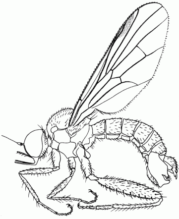 |
| Figure 249. Hybos culiciformis, adult male |
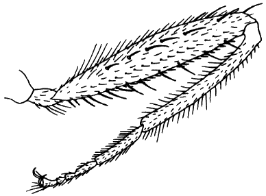 |
| Figure 250. Hybos culiciformis, male hind leg in anterior view. |
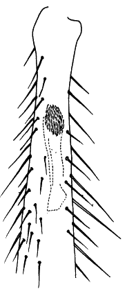 |
| Figure 251. Hybos culiciformis, fore tibia with tubular gland in ventral view. |
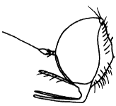 |
| Figure 252. Hybos culiciformis, head in lateral view |
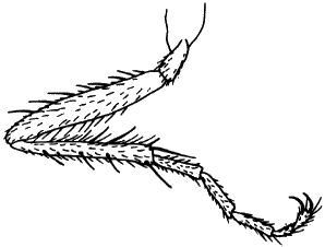 |
| Figure 253. Hybos culiciformis, male fore leg in posterior view. |
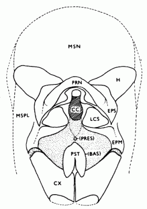 |
| Figure 254. Hybos grossipes, thorax in anterior view. |
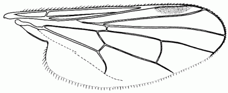 |
| Figure 255. Hybos grossipes, wing |
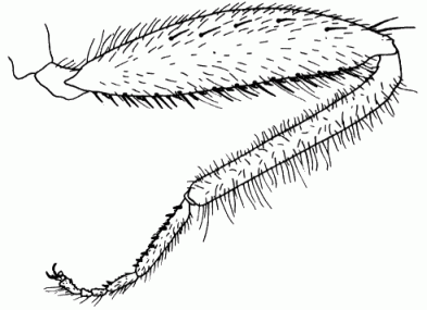 |
| Figure 256. Hybos grossipes, male hind leg in anterior view. |
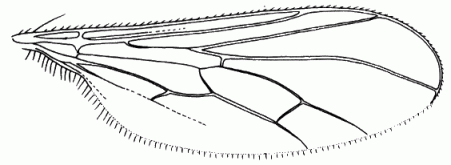 |
| Figure 257. Leptodromiella crassiseta, wing. |
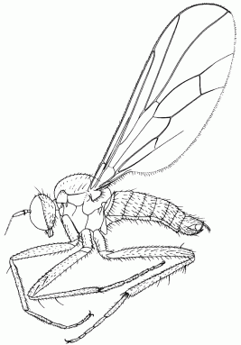 |
| Figure 258. Leptodromiella crassiseta, male in lateral view. |
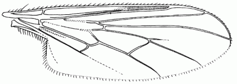 |
| Figure 259. Leptopeza flavipes, wing. |
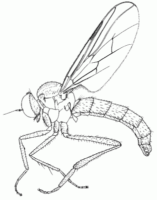 |
| Figure 260. Leptopeza flavipes, male in lateral view. |
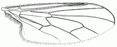 |
| Figure 261. Ocydromia glabricula, wing. |
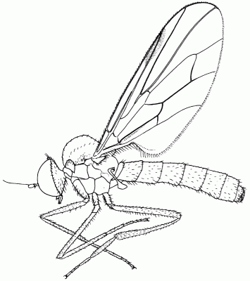 |
| Figure 262. Ocydromia glabricula, male in lateral view. |
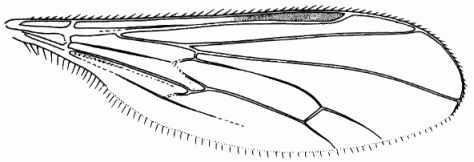 |
| Figure 263. Oropezella sphenoptera, wing. |
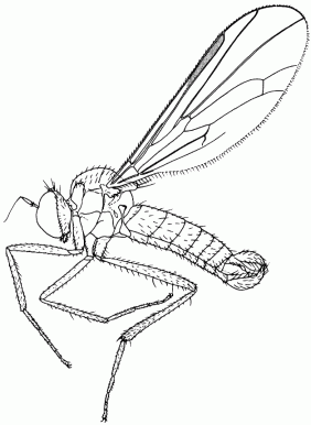 |
| Figure 264. Oropezella sphenoptera, male in lateral view. |
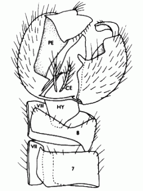 |
| Figure 265. Oropezella sphenoptera, male genitalia in dorsal view (aedeagus omitted). |
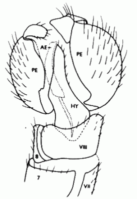 |
| Figure 266. Oropezella sphenoptera, male genitalia in ventral view (aedeagus dotted). |
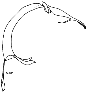 |
| Figure 267. Oropezella sphenoptera, aedeagus. |
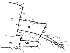 |
| Figure 268. Oropezella sphenoptera, female, tip of abdomen. |
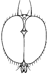 |
| Figure 270. Oropezella sphenoptera, male head in anterior view. |
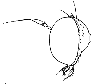 |
| Figure 271. Oropezella sphenoptera, male head in lateral view. |
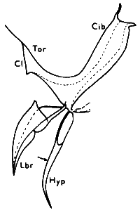 |
| Figure 272. Oropezella sphenoptera, male labrum and hypopharynx in lateral view. |
 |
| Figure 273. Oropezella sphenoptera, male hypopharynx in anterolateral view. |
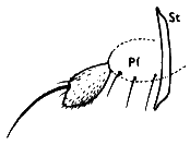 |
| Figure 274. Oropezella sphenoptera, male maxilla and palpus in lateral view. |
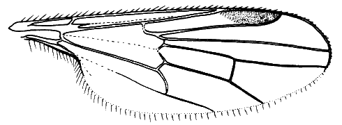 |
| Figure 275. Syndyas nigripes, wing |
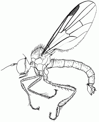 |
| Figure 276. Syndyas ngripes, male in lateral view |
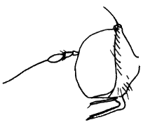 |
| Figure 277. Syndyas nigripes, head in lateral view |
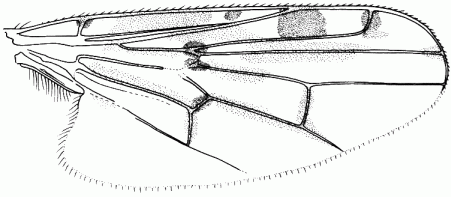 |
| Figure 278. Syneches muscarius, wing |
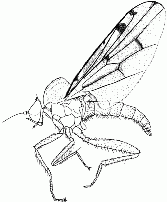 |
| Figure 279. Syneches muscarius, male in lateral view. |
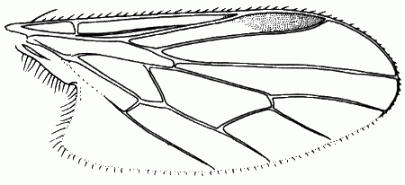 |
| Figure 280. Trichina clavipes, wing. |
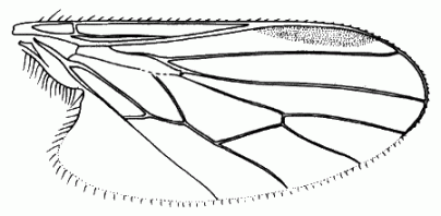 |
| Figure 281. Trichinomyia flavipes, wing |
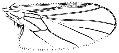 |
| Figure 282. Bicellaria spuria, wing |
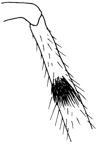 |
| Figure 320. Ocydromia glabricula, male fore tibia with sense organ, anterolateral view. |
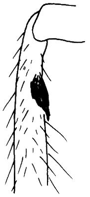 |
| Figure 321. Ocydromia melanopleura, male fore tibia with sense organ, anterolateral view. |
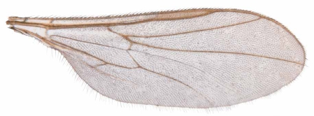 |
| Figure 322. Leptopezella perata, wing. |
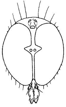 |
| Figure 323. Leptodromiella crassiseta, male head in anterior view. |


