Key: The European species of the genus Stilpon Loew, 1859 (Diptera: Hybotidae)
|
A key to incorporate all European species of the genus Stilpon. The bulk of the keys is adapted from Chvála (1975, 1988) and Collin (1961).
|
| References |
Chvála, M., 1975. The Tachydromiinae (Dipt. Empididae) of Fennoscandia and Denmark. - Fauna Entomologica Scandinavica 3: 1-336.
Chvála, M., 1988. A new species of Stilpon Loew (Dipt., Hybotidae) related to S. nubilus Collin from England and Western Europe. - Entomologist's Monthly Magazine 124: 225-231.
Collin, J.E., 1961. Empididae. - British Flies 6: v-viii, 1-782.
Grootaert, P., & I. Shamshev, 2003. A note on the remarkable fauna (Diptera Empididae Hybotidae Atelestidae) of Corsica. - Bulletin de la Société Royale Belge d'Entomologie 139: 243-252.
Raffone, G., 1994. Descrizione di una specie di Stilpon (Pseudostilpon) Séguy, 1950 de Majorca (Spagna). - Bollettino della Società Entomologica Italiana, Genoa 126: 66-68.
Séguy, E., 1950. Un nouveau genre de Corynétine du Midi de la France (Dipt., Empididae). - Vie et milieu 1: 83-87.
Shamshev, I., & P. Grootaert, 2003. New data on the genus Stilpon Loew (Diptera: Hybotidae) from the Palaearctic region, with description of a new species from Tajikistan. - Belgian Journal of Entomologie 7: 81-86.
Smith, K.G.V., 1965. A new species of Stilpon Loew, 1859 (Diptera: Empididae) from Portugal. - Proceedings of the Royal Entomological Society London, Series B 34: 48-50.
|
| Last modified 16/11/2022 17:16 by Paul Beuk | No of couplets: 10 |
Key (online version)
|
|
1a |
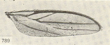
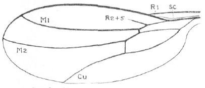
Vein R2+3 abbreviated [Fig. 99].
In S. intermedius occasionally complete. |
|
2 |
| b |
| Vein R2+3 reaching wing margin [Fig. 71]. |
|
4 |
| |
|
2a |
| Wing with more or less parallel anterior and posterior margins [Fig. 150]. |
| S. delamarei (Séguy, 1950) |
|
|
| b |
| Wing more or less ovoid [Fig. 99] [Fig. 151]. |
|
3 |
| |
|
3a |
Vein A2 straight [Fig. 151], third antennal segment subcylindrical.
[The character of vein A2 is not visible when comparing the illustrations of paludosus given by Séguy and of intermedius given by Raffone. Examination of Perris' material will be necessary to illucidate this.] |
| S. paludosus (Perris, 1852) |
|
|
| b |
| Vein A2 curved [Fig. 99], third antennal segment globular. Genitalia as illustrated [Fig. 101] [Fig. 100]. |
| S. intermedius Raffone, 1994 |
|
|
| |
|
4a |
| Halteres with blackish knob, in teneral specimens more brownish. Metatarsi blackish, in teneral specimens brownish. |
| S. gussakovskii Shamshev & Grootaert, 2005 |
|
|
| b |
| Halteres entirely pale. Metatarsi yellow. |
|
5 |
| |
|
5a |
| Wings uniformly faintly yellowish-brown tinged, without a distinct dark pattern [Fig. 71] [Fig. 72]. Preapical bristle beneath fore tibiae indistinct. |
| S. graminum (Fallén, 1815) |
|
|
| b |
| Wings with distinct dark brown clouding at middle, base and tip of wing almost clear [Fig. 73] [Fig. 74]. Fore tibiae with distinct even if small dark preapical bristle beneath . |
|
6 |
| |
|
6a |
| Dark cloud on wing with a clear ovate patch at middle of cell r3. Right cercus of male hypopygium without terminal black spines, at most with some strong black setae [Fig. 118] (left inset). |
|
7 |
| b |
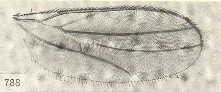
| Wings with a large dark brown cloud at middle, cell r3 without a clear patch [Fig. 73] [Fig. 74]. Male genitalia with long black terminal spines on both cerci [Fig. 70]. |
|
8 |
| |
|
7a |
| Wings rather longer than thorax and abdomen combined, and usually about two and a half times as long as wide, if narrower discal vein more upcurved, and outer crossvein sloping. Genitalia: [Fig. 118]. |
| S. lunatus (Walker, 1851) |
|
|
| b |
| Wings extending very little beyond end of abdomen, and about three times as long as wide. Discal vein straighter and outer crossvein more upright. Genitalia: [Fig. 119]. |
| S. sublunatus Collin, 1961 |
|
|
| |
|
8a |
| Wing without darkened costa [Fig. 148]. Left cercus with three terminal spines [Fig. 146]. |
| S. machadoi Smith, 1965 |
|
|
| b |
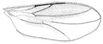
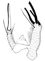
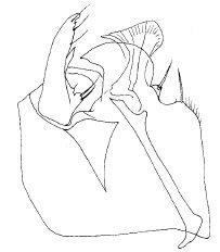
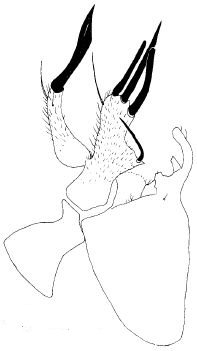
| Wing with costa darkened [Fig. 74] [Fig. 73]. Left cercus with two or three terminal spines [Fig. 149] [Fig. 147] [Fig. 153], if with three, then wings less extensively darkened [Fig. 152]. |
|
9 |
| |
|
9a |
| Wing less extensively darkened [Fig. 152]. Right cercus in male genitalia with three terminal spines [Fig. 153]. |
|
|
| b |
| Wing more extensively darkened [Fig. 73] [Fig. 74]. Right cercus in male genitalia with two terminal spines [Fig. 149] [Fig. 147]. |
|
10 |
| |
|
10a |
| Wings smaller, narrower, and more pointed at the tip; vein R4+5 ends more before the wing tip; brown costal streak is deeper brown and occupies the whole of cell r2+3, right up to the tip of vein R4+5 (rarely the very extreme tip is paler); the lower half of wing beyond vein M is, on the contrary, less clouded [Fig. 73]. Genitalia: right cercus with single apical spine [Fig. 149]. |
| S. nubilus Collin, 1926 |
|
|
| b |
Wing larger and rather rounded at the tip; vein R4+5 ending close to tip of wing; costal streak not as dark with tip of cell r2+3 paler; clouding in posterior half of wing somewhat more extensive [Fig. 74]. Genitalia: right cercus with two apical spines [Fig. 70]
Another three species are know to Chvála (1988):
appediculatus Frey, 1936 (Canary Islands);
undescribed species (Malta);
undescribed species (Rhodos). |
| S. subnubilus Chvála, 1988 |
|
|
| |
| Images |
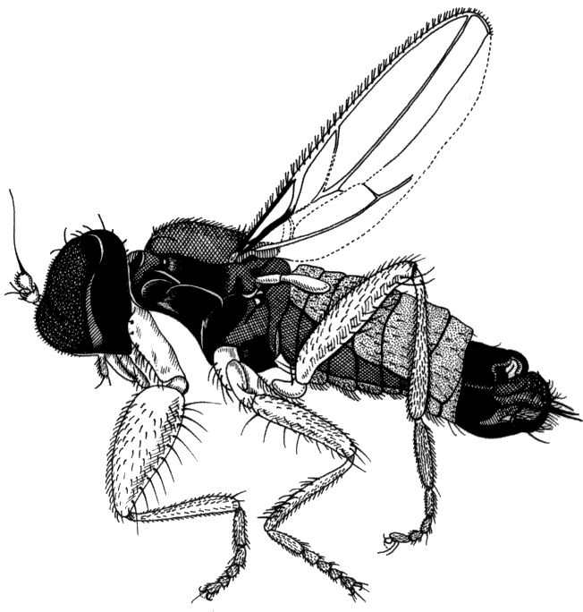 |
| Figure 65. Stilpon graminum, adult male. Size: 1.1-1.3 mm. | (file name: 4_stgrami1.gif) |
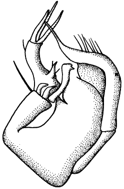 |
| Figure 66. Stilpon graminum, hypopygium with cerci on the left. Right periandrial lamella (x) on the right, left lamella on the left (partly fused with hypandrium). | (file name: 4_stgrami2.gif) |
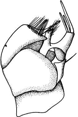 |
| Figure 67. Stilpon graminum, hypopygium with cerci on the right. Right periandrial lamella (x) on the left, cerci on the right. | (file name: 4_stgrami3.gif) |
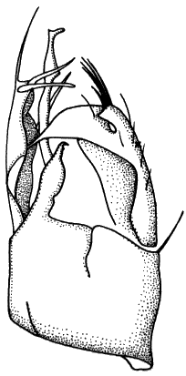 |
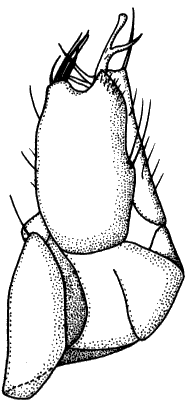
| Figure 68. Stilpon subnubilus, hypopygium with cerci on the left. | (file name: 4_stsubnu2.gif) |
 |
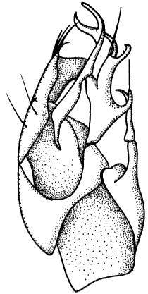
| Figure 69. Stilpon subnubilus, hypopygium with right periandrial lamella in front. | (file name: 4_stsubnu3.gif) |
 |
| Figure 70. Stilpon subnubilus, hypopygium with cerci in front. Right periandrial lamella (x) on the left, left periandrial lamella (partly fused with hypandrium) on the right. | (file name: 4_stsubnu4.gif) |
 |
| Figure 71. Stilpon graminum, wing (macropterous). | (file name: 4_stgrami4.gif) |
 |
| Figure 72. Stilpon graminum, wing (stenopterous). | (file name: 4_stgrami5.gif) |
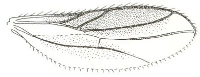 |
| Figure 73. Stilpon nubilus, wing. | (file name: 4_stnubil4.jpg) |
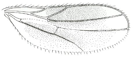 |
| Figure 74. Stilpon subnubilus, wing. | (file name: 4_stsubnu1.jpg) |
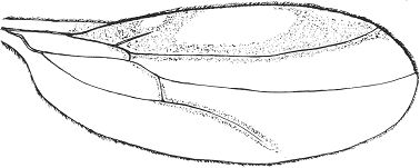 |
| Figure 99. Stilpon intermedius, wing (from Raffone, 1994). | (file name: 4_stinter1.jpg) |
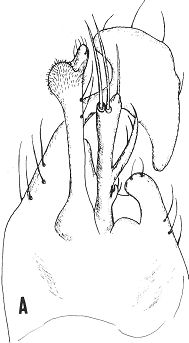 |
| Figure 100. Stilpon intermedius, epandium (from Raffone, 1994). | (file name: 4_stinter2.jpg) |
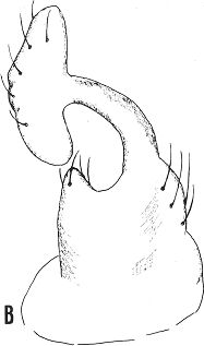 |
| Figure 101. Stilpon intermedius, periandrial lamellae (from Raffone, 1994). | (file name: 4_stinter3.jpg) |
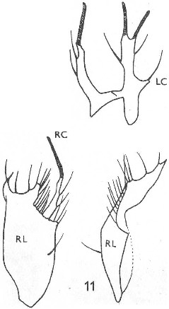 |
| Figure 115. Stilpon nubilus: left; right periandrial lamella with right cercus; right: right periandrial lamella, anterior view. | (file name: 4_stnubil1.jpg) |
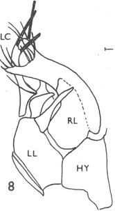 |
| Figure 116. Stilpon nubilus: general view, with cerci on the left. | (file name: 4_stnubil2.jpg) |
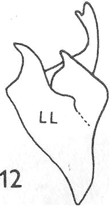 |
| Figure 117. Stilpon nubilus: left periandrial lamella. | (file name: 4_stnubil3.jpg) |
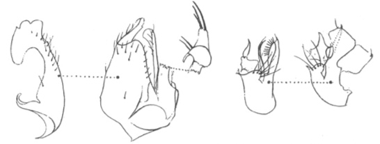 |
| Figure 118. Stilpon lunatus: left: male genitalia: right periandrial lamella (genitalia in ventral view); right: male genitalia: left preiandrial lamella (genitalia in dorsal view). | (file name: 4_stlunat1.jpg) |
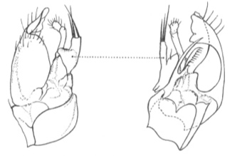 |
| Figure 119. Stilpon sublunatus: left: male genitalia: right periandrial lamella (genitalia in ventral view); right: male genitalia: left preiandrial lamella (genitalia in dorsal view). | (file name: 4_stsublu1.jpg) |
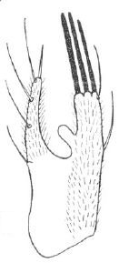 |
| Figure 146. Stilpon machadoi: male cerci. | (file name: 4_stmacha1.jpg) |
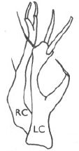 |
| Figure 147. Stilpon subnubilus: male cerci. | (file name: 4_stsubnu5.jpg) |
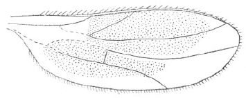 |
| Figure 148. Stilpon machadoi: wing. | (file name: 4_stmacha2.jpg) |
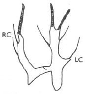 |
| Figure 149. Stilpon nubilus: male cerci. | (file name: 4_stnubil5.jpg) |
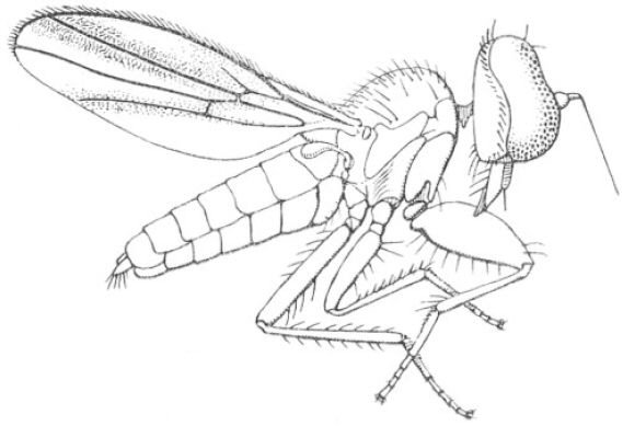 |
| Figure 150. Stilpon delamarei: adult, female. | (file name: 4_stdelam1.jpg) |
 |
| Figure 151. Stilpon paludosus: wing. | (file name: 4_stpalud1.jpg) |
 |
| Figure 152. Stilpon corsicanus: wing. | (file name: 4_stcorsi1.jpg) |
 |
| Figure 153. Stilpon corsicanus: male cerci. | (file name: 4_stcorsi2.jpg) |
 |
| Figure 154. Stilpon corsicanus: male genitalia, ventral view of epandrium. | (file name: 4_stcorsi3.jpg) |
 |
| Figure 155. Stilpon corsicanus: male genitalia, cerci with left epandrial lamella. | (file name: 4_stcorsi4.jpg) |
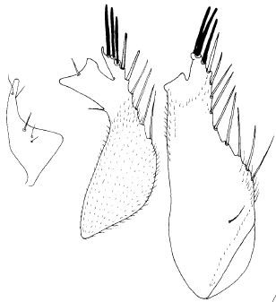 |
| Figure 156. Stilpon corsicanus: male genitalia; left and middle: right epandrial lamella and detail of apex; right: right epandrial lamella in different view. | (file name: 4_stcorsi5.jpg) |


